


 Introduction
Q. What is Human Reproductive System?
Ans: The reproductive events in humans include, Formation of gametes (gametogenesis), i.e., sperms in males and ovum in females, Transfer of sperms into the female genital tract (insemination), Fusion of male and female gametes (fertilisation) leading to formation of zygote. Formation and development of blastocyst and its attachment to the uterine wall (implantation), Embryonic development (gestation) and Delivery of the baby (parturition).
Repr System
- The male reproductive system is located in the pelvis region. It includes a pair of testes along with accessory ducts, glands and the external genitalia.
- The testes are situated outside the abdominal cavity within a pouch called scrotum. The scrotum helps in maintaining the low temperature of the testes (2–2.50C lower than the normal internal body temperature) necessary for spermatogenesis.
- Each testis has about 250 testicular lobules. Each lobule contains one to three highly coiled seminiferous tubules in which sperms are produced.
- Each seminiferous tubule is lined on its inside by two types of cells called male germ cells (spermatogonia) and Sertoli cells.
- The male germ cells undergo meiotic divisions finally leading to sperm formation, while Sertoli cells provide nutrition to the germ cells.
- The regions outside the seminiferous tubules called interstitial spaces, contain small blood vessels and interstitial cells or Leydig cells. Leydig cells synthesise and secrete testicular hormones called androgens [a male sex hormone, such as testosterone. Androgens stimulates or controls the development and maintenance of male characteristics].
- The male sex accessory ducts include rete testis, vasa efferentia, epididymis and vas deferens.
- The seminiferous tubules of the testis open into the vasa efferentia through rete testis.
- The vasa efferentia leave the testis and open into epididymis. The epididymis leads to vas deferens that ascends to the abdomen and loops over the urinary bladder. It receives a duct from seminal vesicle [gland that secrete many of the components of semen] and opens into urethra as the ejaculatory duct. These ducts store and transport the sperms from the testis to the outside through urethra.
- The urethra originates from the urinary bladder and extends through the penis to its external opening called urethral meatus.
- The penis is the male external genitalia. It is made up of special tissue that helps in erection of the penis to facilitate insemination. The enlarged end of penis called the glans penis is covered by a loose fold of skin called foreskin.
- The male accessory glands include paired seminal vesicles, a prostate [releasing a fluid component of semen] and paired bulbourethral glands.
- Secretions of these glands constitute the seminal plasma which is rich in fructose, calcium and certain enzymes.
- The secretions of bulbourethral glands also helps in the lubrication of the penis.
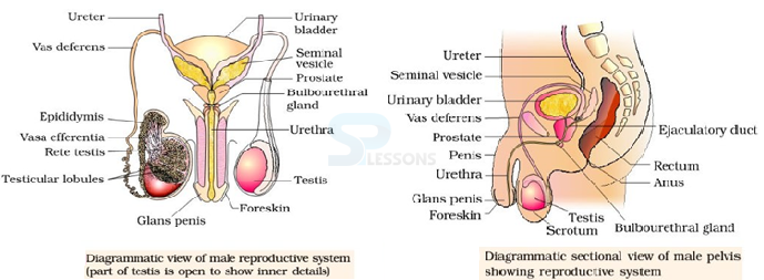
Image: (a) Diagrammatic view of Male reproductive system (b) sectional view of male pelvis
Source: NCERT Text Books
- The female reproductive system consists of a pair of ovaries along with a pair of oviducts, uterus, cervix, vagina and the external genitalia located in pelvic region.
- These parts of the system along with a pair of the mammary glands are integrated structurally and functionally to support the processes of ovulation, fertilisation, pregnancy, birth and child care.
- Ovaries are the primary female sex organs [testis in males] that produce the female gamete (ovum) [sperm in males] and several steroid hormones (ovarian hormones).
- The ovaries are located one on each side of the lower abdomen. Each ovary is connected to the pelvic wall and uterus by ligaments.
- Each ovary is covered by a thin epithelium which encloses the ovarian stroma. The stroma is divided into two zones – a peripheral cortex and an inner medulla.
- The oviducts (fallopian tubes), uterus and vagina constitute the female accessory ducts.
- Each fallopian tube extends from the periphery of each ovary to the uterus, the part closer to the ovary is the funnel-shaped infundibulum.
- The edges of the infundibulum possess finger-like projections called fimbriae, which help in collection of the ovum after ovulation. The infundibulum leads to a wider part of the oviduct called ampulla.
- The last part of the oviduct, isthmus has a narrow lumen and it joins the uterus. The uterus is single and it is also called womb. The shape of the uterus is like an inverted pear.
- It is supported by ligaments attached to the pelvic wall. The uterus opens into vagina through a narrow cervix. The cavity of the cervix is called cervical canal which along with vagina forms the birth canal.
- The wall of the uterus has three layers of tissue. The external thin membranous perimetrium, middle thick layer of smooth muscle, myometrium and inner glandular layer called endometrium that lines the uterine cavity.
- The endometrium undergoes cyclical changes during menstrual cycle while the myometrium exhibits strong contraction during delivery of the baby.
- The female external genitalia include mons pubis, labia majora, labia minora, hymen and clitoris.
- Mons pubis is a cushion of fatty tissue covered by skin and pubic hair. The labia majora are fleshy folds of tissue, which extend down from the mons pubis and surround the vaginal opening.
- Thelabia minora are paired folds of tissue under the labia majora. The opening of the vagina is often covered partially by a membrane called hymen.
- The clitoris is a tiny finger-like structure which lies at the upper junction of the two labia minora above the urethral opening.
- The hymen is often torn during the first coitus (intercourse). However, it can also be broken by a sudden fall or jolt, insertion of a vaginal tampon, active participation in some sports like horseback riding, cycling, etc.
- In some women the hymen persists even after coitus. In fact, the presence or absence of hymen is not a reliable indicator of virginity or sexual experience.
- A functional mammary gland is characteristic of all female mammals. The mammary glands are paired structures (breasts) that contain glandular tissue and variable amount of fat.
- The glandular tissue of each breast is divided into 15-20 mammary lobes containing clusters of cells called alveoli. The cells of alveoli secrete milk, which is stored in the cavities (lumens) of alveoli. The alveoli open into mammary tubules.
- The tubules of each lobe join to form a mammary duct. Several mammary ducts join to form a wider mammary ampulla which is connected to lactiferous duct through which milk is sucked out.
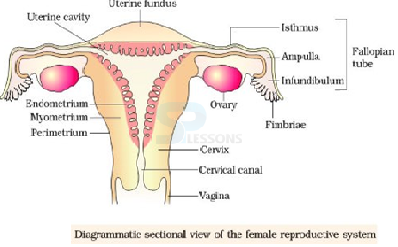
Image: Female Reproductive system
Source: NCERT Text Books
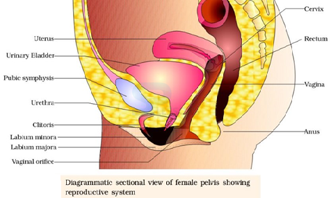
Image: Female pelvis showing reproductive system
Source: NCERT Text Books
- The primary sex organs – the testis in the males and the ovaries in the females produce gametes, i.e, sperms and ovum, respectively, by the process called gametogenesis.
- In testis, the immature male germ cells (spermatogonia) produce sperms by spermatogenesis that begins at puberty.
- The spermatogonia (sing. spermatogonium) present on the inside wall of seminiferous tubules multiply by mitotic division and increase in numbers. Each spermatogonium is diploid and contains 46 chromosomes.
- Some of the spermatogonia called primary spermatocytes periodically undergo meiosis.
- A primary spermatocyte completes the first meiotic division (reduction division) leading to formation of two equal, haploid cells called secondary spermatocytes, which have only 23 chromosomes each.
- The secondary spermatocytes undergo the second meiotic division to produce four equal, haploid spermatids.
- The spermatids are transformed into spermatozoa (sperms) by the process called spermiogenesis. After spermiogenesis, sperm heads become embedded in the Sertoli cells, and are finally released from the seminiferous tubules by the process called spermiation.
- Spermatogenesis starts at the age of puberty due to significant increase in the secretion of gonadotropin releasing hormone (GnRH). This, if you recall, is a hypothalamic hormone.
- The increased levels of GnRH then acts at the anterior pituitary gland and stimulates secretion of two gonadotropins – luteinising hormone (LH) and follicle stimulating hormone (FSH).
- LH acts at the Leydig cells and stimulates synthesis and secretion of androgens. Androgens, in turn, stimulate the process of spermatogenesis.
- FSH acts on the Sertoli cells and stimulates secretion of some factors which help in the process of spermiogenesis.
- Sperm is a microscopic structure composed of a head, neck, a middle piece and a tail. A plasma membrane envelops the whole body of sperm.
- The sperm head contains an elongated haploid nucleus, the anterior portion of which is covered by a cap-like structure, acrosome. The acrosome is filled with enzymes that help fertilization of the ovum.
- The middle piece possesses numerous mitochondria, which produce energy for the movement of tail that facilitate sperm motility essential for fertilization.
- The human male ejaculates about 200 to 300 million sperms during a coitus of which, for normal fertility, at least 60 percent sperms must have normal shape and size and at least 40 per cent of them must show vigorous motility.
- Sperms released from the seminiferous tubules, are transported by the accessory ducts.
- Secretions of epididymis, vas deferens, seminal vesicle and prostate are essential for maturation and motility of sperms.
- The seminal plasma along with the sperms constitute the semen. The functions of male sex accessory ducts and glands are maintained by the testicular hormones (androgens).
- The process of formation of a mature female gamete is called oogenesis which is markedly different from spermatogenesis.
- Oogenesis is initiated during the embryonic development stage when a couple of million gamete mother cells (oogonia) are formed within each fetal ovary; no more oogonia are formed and added after birth.
- These cells start division and enter into prophase-I of the meiotic division and get temporarily arrested at that stage, called primary oocytes.
- Each primary oocyte then gets surrounded by a layer of granulosa cells and is called the primary follicle.
- A large number of these follicles degenerate during the phase from birth to puberty. Therefore, at puberty only 60,000-80,000 primary follicles are left in each ovary.
- The primary follicles get surrounded by more layers of granulosa cells and a new theca and are called secondary follicles. The secondary follicle soon transforms into a tertiary follicle which is characterised by a fluid filled cavity called antrum.
- At this stage the primary oocyte within the tertiary follicle grows in size and completes its first meiotic division. It is an unequal division resulting in the formation of a large haploid secondary oocyte and a tiny first polar body.
- The secondary oocyte retains bulk of the nutrient rich cytoplasm of the primary oocyte.
- The tertiary follicle further changes into the mature follicle or Graafian follicle. The secondary oocyte forms a new membrane called zona pellucida surrounding it.
- The Graafian follicle now ruptures to release the secondary oocyte (ovum) from the ovary by the process called ovulation.
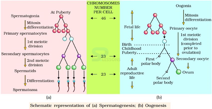
Image: Representation of spermatogenesis and oogenesis
Source: NCERT Text Books
What would be the number of chromosome in the spermatids? 23 chromosomes.- The reproductive cycle in the female primates (e.g. monkeys, apes and human beings) is called menstrual cycle. The first menstruation begins at puberty and is called menarche.
- In human females, menstruation is repeated at an average interval of about 28/29 days, and the cycle of events starting from one menstruation till the next one is called the menstrual cycle.
- One ovum is released (ovulation) during the middle of each menstrual cycle. The cycle starts with the menstrual phase, when menstrual flow occurs and it lasts for 3-5 days.
- The menstrual flow results due to breakdown of endometrial lining of the uterus and its blood vessels which forms liquid that comes out through vagina. Menstruation only occurs if the released ovum is not fertilized.
- Lack of menstruation may be indicative of pregnancy. However, it may also be caused due to some other underlying causes like stress, poor health etc.
- The menstrual phase is followed by the follicular phase. During this phase, the primary follicles in the ovary grow to become a fully mature Graafian follicle and simultaneously the endometrium of uterus regenerates through proliferation.
- These changes in the ovary and the uterus are induced by changes in the levels of pituitary and ovarian hormones.
- The secretion of gonadotropins (LH and FSH) increases gradually during the follicular phase, and stimulates follicular development as well as secretion of estrogens by the growing follicles.
- Both LH and FSH attain a peak level in the middle of cycle (about 14 th day). Rapid secretion of LH leading to its maximum level during the mid-cycle called LH surge induces rupture of Graafian follicle and thereby the release of ovum (ovulation).
- The ovulation (ovulatory phase) is followed by the luteal phase during which the remaining parts of the Graafian follicle transform as the corpus luteum.
- The corpus luteum secretes large amounts of progesterone which is essential for maintenance of the endometrium. Such an endometrium is necessary for implantation of the fertilised ovum and other events of pregnancy.
- During pregnancy all events of the menstrual cycle stop and there is no menstruation. In the absence of fertilisation, the corpus luteum degenerates. This causes disintegration of the endometrium leading to menstruation, marking a new cycle.
- In human beings, menstrual cycles ceases around 50 years of age; that is termed as menopause.
- Cyclic menstruation is an indicator of normal reproductive phase and extends between menarche and menopause. Fertilisation And Implantation
- During copulation (coitus) semen is released by the penis into the vagina (insemination). The motile sperms swim rapidly, pass through the cervix, enter into the uterus and finally reach the ampullary region of the fallopian tube.
- The ovum released by the ovary is also transported to the ampullary region where fertilisation takes place.
- Fertilisation can only occur if the ovum and sperms are transported simultaneously to the ampullary region. This is the reason why not all copulations lead to fertilisation and pregnancy.
- The process of fusion of a sperm with an ovum is called fertilisation. During fertilisation, a sperm comes in contact with the zona pellucida layer of the ovum and induces changes in the membrane that block the entry of additional sperms. Thus, it ensures that only one sperm can fertilise an ovum.
- The secretions of the acrosome help the sperm enter into the cytoplasm of the ovum through the zona pellucida and the plasma.
- Ovum surrounded by few sperm blastomeres is called a morula. The morula continues to divide and transforms into blastocyst as it moves further into the uterus.
- The blastomeres in the blastocyst are arranged into an outer layer called trophoblast and an inner group of cells attached to trophoblast called the inner cell mass. The trophoblast layer then gets attached to the endometrium and the inner cell mass gets differentiated as the embryo.
- After attachment, the uterine cells divide rapidly and covers the blastocyst. As a result, the blastocyst becomes embedded in the endometrium of the uterus. This is called implantation and it leads to pregnancy.
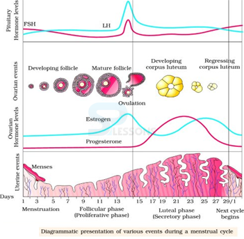
Image: Diagrammatic representation of menstrual cycle
Source: NCERT Text Books
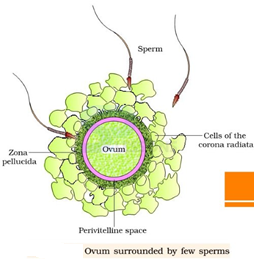
Image: ovum surrounded by few sperms
Source: NCERT Text Books
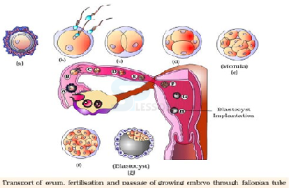
Image: Transport of ovum
Source: NCERT Text Books
- Have you heard of test-tube babies? In some women, oviducts are blocked. These women are unable to bear babies because sperms cannot reach the egg for fertilization. In such cases, doctors collect freshly released egg and sperms and keep them together for a few hours for IVF or In Vitro Fertilization (fertilization outside the body).
- In case fertilization occurs, the zygote is allowed to develop for about a week and then it is placed in the mother’s uterus. Complete development takes place in the uterus and the baby is born like any other baby.
- Babies born through this technique are called test-tube babies. This term is actually misleading because babies cannot grow in test tubes.
- After implantation, finger-like projections appear on the trophoblast called chorionic villi which are surrounded by the uterine tissue and maternal blood.
- The chorionic villi and uterine tissue become interdigitated with each other and jointly form a structural and functional unit between developing embryo (foetus) and maternal body called placenta.
- The placenta facilitate the supply of oxygen and nutrients to the embryo and also removal of carbon dioxide and excretory/waste materials produced by the embryo.
- The placenta is connected to the embryo through an umbilical cord which helps in the transport of substances to and from the embryo.
- Placenta also acts as an endocrine tissue and produces several hormones like human chorionic gonadotropin (hCG), human placental lactogen (hPL), estrogens, progestogens, etc.
- In the later phase of pregnancy, a hormone called relaxin is also secreted by the ovary. Let us remember that hCG, hPL and relaxin are produced in women only during pregnancy.
- In addition, during pregnancy the levels of other hormones like estrogens, progestogens, cortisol, prolactin, thyroxine, etc., are increased several folds in the maternal blood.
- Increased production of these hormones is essential for supporting the fetal growth, metabolic changes in the mother and maintenance of pregnancy.
- Immediately after implantation, the inner cell mass (embryo) differentiates into an outer layer calledectoderm and an inner layer called endoderm. A mesoderm soon appears between the ectoderm and the endoderm [triploblastic]. These three layers give rise to all tissues (organs) in adults.
- It needs to be mentioned here that the inner cell mass contains certain cells called stem cells which have the potency to give rise to all the tissues and organs.
- The human pregnancy lasts 9 months. In human beings, after one month of pregnancy, the embryo’s heart is formed. The first sign of growing foetus may be noticed by listening to the heart sound carefully through the stethoscope.
- By the end of the second month of pregnancy, the foetus develops limbs and digits. By the end of 12 weeks (first trimester), most of the major organ systems are formed, for example, the limbs and external genital organs are well developed.
- The first movements of the foetus and appearance of hair on the head are usually observed during the fifth month. By the end of about 24 weeks (end of second trimester), the body is covered with fine hair, eye-lids separate, and eyelashes are formed. By the end of nine months of pregnancy, the foetus is fully developed and is ready for delivery.
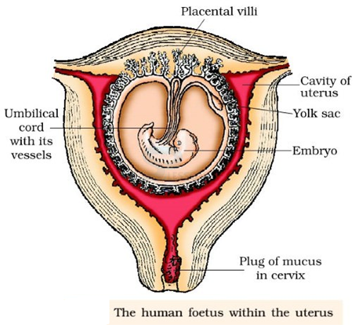
Image: human foetus within the uterus
Source: NCERT Text Books
- The average duration of human pregnancy is about 9 months which is called the gestation period. Vigorous contraction of the uterus at the end of pregnancy causes expulsion/delivery of the fetus. This process of delivery of the fetus (childbirth) is called parturition.
- Parturition is induced by a complex neuroendocrine mechanism. The signals for parturition originate from the fully developed fetus and the placenta which induce mild uterine contractions called foetal ejection reflex. This triggers the release of oxytocin from the maternal pituitary.
- Oxytocin acts on the uterine muscle and causes stronger uterine contractions, which in turn stimulates the further secretion of oxytocin. The stimulatory reflex between the uterine contraction and oxytocin secretion continues resulting in stronger and stronger contractions. This leads to the expulsion of the baby out of the uterus through the birth canal – parturition.
- Soon after the infant is delivered, the placenta is also expelled out of the uterus. The mammary glands of the female undergo differentiation during pregnancy and start producing milk towards the end of pregnancy by the process called lactation. This helps the mother in feeding the newborn.
- The milk produced during the initial few days of lactation is called colostrum which contains several antibodies absolutely essential to develop resistance for the new-born babies.
- Breast-feeding during the initial period of infant growth is recommended by doctors for bringing up a healthy baby.
Biology - Related Information
Biotechnology
General Science Practice Set
Important General Science Quiz
General Science - Biology - DNA RNA
