


 Introduction
Q. What are Human Movements?
Ans: Types of Movements
Voluntary movements are called locomotion. Walking, running, climbing, flying, swimming are all some forms of locomotory movements. Cells of the human body exhibit three main types of movements, namely, amoeboid, ciliary and muscular. Some specialized cells in our body like macrophages and leucocytes in blood exhibit amoeboid movement. It is effected by pseudopodia formed by the streaming of protoplasm .
Key Points
- Ciliary movement occurs in most of our internal tubular organs which are lined by ciliated epithelium.
- The coordinated movements of cilia in the trachea help us in removing dust particles and some of the foreign substances inhaled along with the atmospheric air.
- Passage of ova through the female reproductive tract is also facilitated by the ciliary movement.
- Movement of our limbs, jaws, tongue, etc. require muscular movement. The contractile property of muscles are effectively used for locomotion and other movements by human beings and majority of multicellular organisms. Locomotion requires a perfect coordinated activity of muscular, skeletal and neural systems.
Movements
- Muscle is a specialized tissue of mesodermal origin.
- About 40-50 percent of the body weight of a human adult is contributed by muscles.
- They have special properties like excitability, contractility, extensibility, and elasticity.
- Muscles have been classified using different criteria, namely location, appearance and nature of regulation of their activities. Based on their location, three types of muscles are identified : (i) Skeletal (ii) Visceral and (iii) Cardiac.
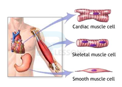
Image: Muscle types
Source: NCERT Text Books
- Skeletal muscles are closely associated with the skeletal components of the body. They have a striped appearance under the microscope and hence are called striated muscles.
- As their activities are under the voluntary control of the nervous system, they are known as voluntary muscles They are primarily involved in locomotory actions and changes of body postures.
- Each organized skeletal muscle in our body is made of a number of muscle bundles or fascicles held together by a common connective tissue layer called fascia.
- Each muscle bundle contains a number of muscle fibres. Each muscle fibre is lined by the plasma membrane called sarcolemma enclosing the sarcoplasm.
- The endoplasmic reticulum, i.e., sarcoplasmic reticulum of the muscle fibres is the store house of calcium ions.
- A characteristic feature of the muscle fibre is the presence of a large number of parallelly arranged filaments in the sarcoplasm called myofilaments or myofibrils.
- Each myofibril has alternate dark and light bands on it. The striated appearance is due to the distribution pattern of two important proteins - Actin and Myosin.
- Actin and myosin are polymerized proteins with contractility. A motor neuron carries signal to the muscle fibre which generates an action potential in it. This causes the release of Ca++ from sarcoplasmic reticulum.
- Ca++ activates actin which binds to the myosin head to form a cross bridge. These cross bridges pull the actin filaments causing them to slide over the myosin filaments and thereby causing contraction. Ca++ are then returned to sarcoplasmic reticulum which inactivate the actin. Cross bridges are broken and the muscles relax.
- Muscles are classified as Red and White fibres based primarily on the amount of red coloured myoglobin pigment in them.
- Visceral muscles are located in the inner walls of hollow visceral organs of the body like the alimentary canal, reproductive tract, etc.
- They do not exhibit any striation and are smooth in appearance. Hence, they are called smooth muscles (nonstriated muscle).
- Their activities are not under the voluntary control of the nervous system and are therefore known as involuntary muscles.
- They assist, for example, in the transportation of food through the digestive tract and gametes through the genital tract.
- As the name suggests, Cardiac muscles are the muscles of heart. Many cardiac muscle cells assemble in a branching pattern to form a cardiac muscle.
- Based on appearance, cardiac muscles are striated. They are involuntary in nature as the nervous system does not control their activities directly.
- Skeletal system consists of a framework of bones and a few cartilages. Bone and cartilage are specialized connective tissues.
- The former has a very hard matrix due to calcium salts in it and the latter has slightly pliable matrix due to chondroitin salts.
- In human beings, this system is made up of 206 bones and a few cartilages. It is grouped into two principal divisions - the axial and the appendicular skeleton.
- Axial skeleton comprises 80 bones distributed along the main axis of the body. The skull, vertebral column, sternum, and ribs constitute axial skeleton. Joints
- Joints are essential for all types of movements involving the bony parts of the body. Locomotory movements are no exception to this.
- Joints are points of contact between bones, or between bones and cartilages.
- Joints have been classified into three major structural forms, namely, fibrous, cartilaginous and synovial.
- Fibrous joints do not allow any movement. This type of joint is shown by the flat skull bones which fuse end-to-end with the help of dense fibrous connective tissues in the form of sutures, to form the cranium.
- In cartilaginous joints, the bones involved are joined together with the help of cartilages. The joint between the adjacent vertebrae in the vertebral column is of this pattern and it permits limited movements.
- Synovial joints are characterized by the presence of a fluid-filled synovial cavity between the articulating surfaces of the two bones. Such an arrangement allows considerable movement. These joints help in locomotion and many other movements.
- Ball and socket joint, hinge joint (knee joint), pivot joint, gliding joint and saddle joint are some examples.
- 1. Ball and socket joints
2. Pivotal Joint: The joint where our neck joins the head is a pivotal joint.
3. Hinge joints
4. Fixed joints
- There are some bones in our head that are joined together at some joints. The bones cannot move at these joints. Such joints are called fixed joints.
- When you open your mouth wide, you can move your lower jaw away from your head, isn’t it? Try to move your upper jaw, now. Are you able to move it? There is a joint between the upper jaw and the rest of the head which is a fixed joint.
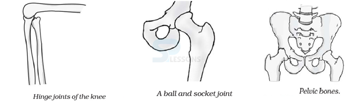
Image: (a)Hinge joints of knee (b)A ball and the socket joint (c)pelvic bones
Source: NCERT Text Books
- Arthritis: Inflammation of joints.
- Osteoporosis: Age-related disorder characterized by decreased bone mass and increased chances of fractures. Decreased levels of estrogen is a common cause.
- Gout: Inflammation of joints due to accumulation of uric acid crystals.
Biology - Related Information
General Science Facts
General Science Practice Set
Important General Science Quiz
General Science - Biology - DNA RNA
